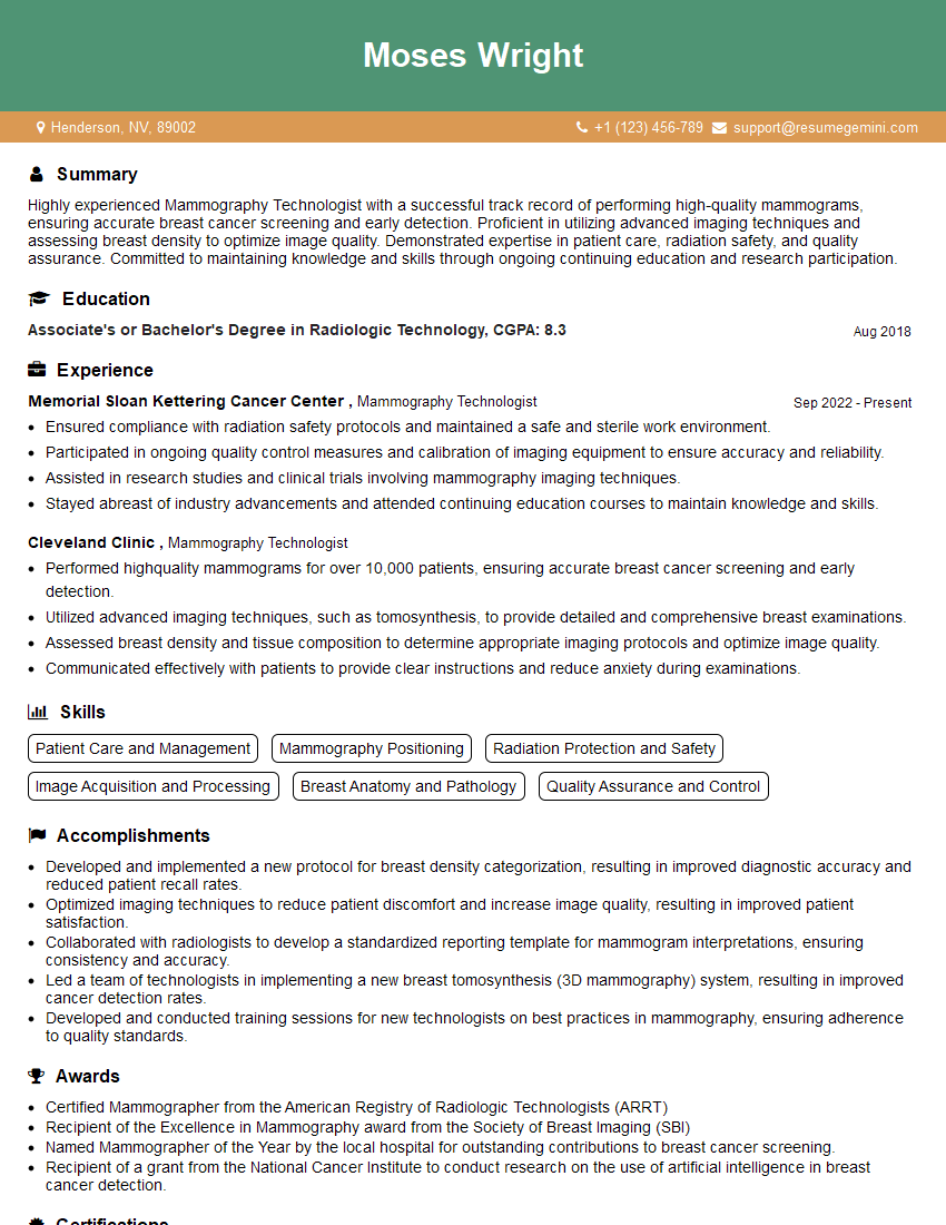
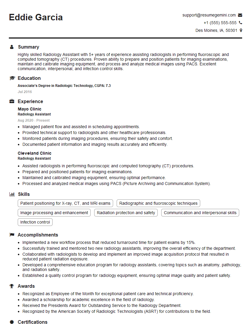
Interviews are more than just a Q&A session—they’re a chance to prove your worth. This blog dives into essential Radiology Technician interview questions and expert tips to help you align your answers with what hiring managers are looking for. Start preparing to shine!
Questions Asked in Radiology Technician Interview
Q 1. What are the different types of ionizing radiation used in radiology?
In radiology, we primarily utilize ionizing radiation to create medical images. Ionizing radiation carries enough energy to remove electrons from atoms, leading to ionization. This ionization process is what allows us to capture the image data. The main types used are:
- X-rays: These are electromagnetic waves with high energy. They are produced by an X-ray tube and are widely used in various imaging modalities, including radiography (conventional X-rays), fluoroscopy (live X-ray imaging), and computed tomography (CT).
- Gamma rays: These are also electromagnetic waves, but they originate from radioactive isotopes. Gamma rays are used in nuclear medicine imaging techniques such as single-photon emission computed tomography (SPECT) and positron emission tomography (PET).
The choice of radiation type depends on the specific imaging modality and the information needed. For instance, X-rays are excellent for visualizing bone density, while gamma rays can help detect the metabolic activity of tissues.
Q 2. Explain the ALARA principle in radiology.
ALARA stands for “As Low As Reasonably Achievable.” It’s a fundamental principle in radiation protection, emphasizing that radiation exposure should be minimized whenever possible, without compromising the diagnostic quality of the image. Think of it as a balance: we want to get the best possible image for the patient’s diagnosis, but we need to do so with the lowest possible radiation dose.
To achieve ALARA, we employ various strategies, including optimizing technical factors (like kilovoltage and milliamperage in X-ray), using appropriate shielding (lead aprons, thyroid shields), and employing image processing techniques to reduce noise and improve image quality. For example, if we need a chest X-ray, using the lowest possible mAs while still getting a clear image reduces the patient’s dose. Proper patient positioning is also critical to ALARA, as it ensures that the target area receives the appropriate radiation dose, minimizing unnecessary exposure to other areas.
Q 3. Describe the process of performing a chest X-ray.
Performing a chest X-ray is a relatively straightforward procedure, but precision is key. The steps generally involve:
- Patient Preparation: The patient removes any metal objects that could interfere with the image (jewelry, etc.) and is instructed to remove their top clothing to expose the chest area.
- Positioning: The patient stands or sits facing the X-ray machine, with their chest against the image receptor. Proper positioning ensures a clear and unobstructed view of the lungs and heart. We carefully align the patient to minimize rotation and ensure the heart is centered.
- Image Acquisition: The radiographer adjusts the machine’s settings (kVp, mAs) based on the patient’s size and the desired image quality. The exposure is made, typically with the patient holding their breath to reduce motion blur. The exposure time is very short.
- Image Review: After the exposure, the image is quickly reviewed to ensure proper penetration, positioning, and overall image quality. If necessary, repeat images may be taken.
The entire process emphasizes patient comfort and safety, alongside technical precision to acquire a diagnostic image.
Q 4. What are the safety precautions for operating an X-ray machine?
Safety is paramount in radiology. Precautions include:
- Radiation Shielding: Lead aprons and thyroid shields are used to protect the radiographer and other personnel from scattered radiation. We always stand behind a protective barrier when possible during exposure.
- Distance: The inverse square law dictates that radiation intensity decreases rapidly with increasing distance from the source. We maintain a safe distance from the X-ray tube during exposure.
- Time Minimization: Keeping exposure times as short as possible reduces radiation dose to both the patient and the technologist. This requires efficient workflows and well-trained staff.
- Proper Equipment Maintenance: Regular servicing and calibration of X-ray machines are essential to ensure they operate safely and efficiently.
- Personal Monitoring: Many radiology professionals wear dosimeters that measure their cumulative radiation exposure, ensuring their safety within regulatory limits.
Adherence to these precautions is crucial in minimizing occupational exposure to ionizing radiation.
Q 5. How do you ensure proper patient positioning for different imaging modalities?
Proper patient positioning is crucial for obtaining high-quality images in all imaging modalities. It’s both an art and a science, requiring a thorough understanding of anatomy and the principles of each modality. Different projections and positions are necessary to visualize different structures, minimizing superimposition and artifacts.
For example, in chest X-rays, we use precise positioning to ensure that the heart is centrally located and that the lungs are fully expanded. In CT scans, we meticulously plan the scan parameters to ensure the entire area of interest is covered and artifacts are minimized. MRI requires extremely precise positioning to minimize motion artifacts. We use anatomical landmarks (e.g., spinous processes, acromioclavicular joints) and imaging devices to help ensure correct positioning. Thorough training and experience are critical for accurate patient positioning in radiology.
Q 6. What are the common artifacts seen in CT scans and how are they addressed?
CT scans, while powerful, are susceptible to various artifacts that can degrade image quality and impede diagnosis. Common artifacts include:
- Motion artifacts: These are caused by patient movement during the scan, resulting in blurring or streaking in the image. Addressing this involves using breath-hold techniques, sedation for uncooperative patients, or faster scan times.
- Metal artifacts: Metallic implants (e.g., hip replacements) can cause streaking and obscuring of underlying structures. Specialized scanning techniques or image processing algorithms can often mitigate these artifacts.
- Beam hardening artifacts: This occurs when the X-ray beam passes through dense structures, leading to a cupping or streak artifact. Using different algorithms or adjusting technical parameters can help address it.
- Ring artifacts: These appear as circular lines in the image and can be caused by detector malfunction or calibration issues. This typically requires technical adjustment or repair of the CT machine.
Identifying and understanding the cause of an artifact is essential for selecting the appropriate solution, whether it’s adjusting the scan parameters, employing post-processing techniques, or even rescanning the patient.
Q 7. Explain the difference between T1 and T2 weighted MRI images.
T1-weighted and T2-weighted MRI images are both fundamental sequences used in MRI, but they provide different tissue contrast. This is because they differ in the time point at which the signal is measured in the MRI sequence.
T1-weighted images show good anatomical detail. Fat appears bright, while water appears dark. This is why T1-weighted images are often used to visualize anatomy and assess overall structure.
T2-weighted images are more sensitive to water content. Water and edema appear bright, while fat appears dark. T2-weighted images are therefore very useful for detecting pathologies involving fluid, such as inflammation, edema, or tumors.
Choosing between T1 and T2 weighted images depends entirely on the clinical question. For example, if a clinician is looking for a brain tumor, T2-weighted images may be more helpful because of their sensitivity to edema, a characteristic feature of many tumors. However, if they are assessing the morphology of the tumor, a T1-weighted sequence may be preferred for its anatomical detail.
Q 8. What are the contraindications for MRI scans?
MRI scans use strong magnetic fields and radio waves to create detailed images of the body’s internal structures. However, several contraindications exist, meaning certain conditions make an MRI scan unsafe or inappropriate. These include:
- Presence of metallic implants or devices: Ferromagnetic materials (strongly attracted to magnets) like some aneurysm clips, pacemakers, and certain types of shrapnel can be dislodged or malfunction during an MRI scan, posing serious risks. For example, a pacemaker could be damaged, leading to life-threatening arrhythmias.
- Claustrophobia: The confined space of the MRI machine can trigger severe anxiety or panic attacks in claustrophobic individuals. Alternative imaging methods might be considered.
- Certain medical devices: Some implanted devices, even if not ferromagnetic, can be affected by the MRI’s strong magnetic field, potentially leading to malfunction or inaccurate readings. These might include insulin pumps or cochlear implants – careful assessment is necessary.
- Unstable medical conditions: Patients with certain unstable medical conditions, such as those requiring constant monitoring, may not be suitable for an MRI scan, as it may prolong their time away from crucial monitoring equipment.
- Pregnancy: While generally considered safe, MRI scans during pregnancy are often avoided unless absolutely necessary due to the lack of extensive long-term research on fetal effects.
- Kidney disease (with gadolinium-based contrast): Patients with severe kidney impairment should avoid gadolinium-based contrast agents, as it can accumulate in the body, leading to nephrogenic systemic fibrosis (NSF), a rare but potentially serious condition.
It’s crucial to thoroughly screen patients before scheduling an MRI to identify and mitigate potential risks. A detailed medical history and a discussion of implanted devices are essential parts of this process.
Q 9. Describe the process of performing a mammogram.
A mammogram is an X-ray examination of the breasts used to detect breast cancer and other breast abnormalities. The process involves these steps:
- Patient positioning: The patient is positioned at the mammographic unit, typically with their breasts compressed between two plates. Compression is crucial for several reasons: it reduces the thickness of the breast tissue, allowing for clearer images with less radiation; it helps spread out the breast tissue, reducing image overlap; and it helps to hold the breast still, reducing motion blur. The level of compression is adjusted to ensure patient comfort while still providing optimal image quality.
- Image acquisition: Low-dose X-rays are used to capture images from different angles of each breast (typically two views per breast: craniocaudal and mediolateral oblique). The technologist carefully positions the breast to ensure proper image orientation and minimize artifacts.
- Image review: The radiologist reviews the mammograms for any abnormalities. Areas of concern might lead to further imaging, such as ultrasound or biopsy.
It’s important to explain the procedure thoroughly to the patient, answering any questions and addressing concerns about compression discomfort. Patient communication and a calm, reassuring demeanor are key to making the experience as comfortable as possible. Mammography is a vital tool for early breast cancer detection, and patient education plays a critical role in ensuring compliance and reducing anxiety.
Q 10. What are the safety measures for patients undergoing fluoroscopy?
Fluoroscopy is a dynamic imaging technique that uses X-rays to provide real-time visualization of internal structures during procedures. Patient safety is paramount and requires strict adherence to ALARA principles (As Low As Reasonably Achievable). This includes:
- Minimizing exposure time: The fluoroscopy beam should only be activated when absolutely necessary and for the shortest duration possible. Using pulsed fluoroscopy (intermittent X-ray pulses) rather than continuous fluoroscopy significantly reduces radiation exposure.
- Optimizing image quality: Using appropriate kilovoltage (kVp) and milliamperage (mA) settings ensures good image quality with the lowest possible radiation dose. Proper collimation (restricting the X-ray beam to the area of interest) also reduces unnecessary radiation to surrounding tissues.
- Shielding: Using lead aprons and other protective shielding for both the patient and the technologist reduces radiation exposure to non-target areas. This is particularly important for the reproductive organs.
- Distance: Maintaining a safe distance from the X-ray source reduces radiation exposure. The inverse square law highlights the importance of distance in radiation protection.
- Monitoring radiation dose: Radiation dose monitors are used to track the radiation dose received by the patient and the technologist. This helps to ensure that ALARA principles are followed and that exposure remains within safe limits.
For example, during a barium swallow, the technologist will only activate the fluoroscopy beam during the actual swallow, minimizing the patient’s exposure. Consistent adherence to these safety measures is crucial in minimizing the risks associated with fluoroscopy.
Q 11. How do you handle a patient who is claustrophobic during an MRI scan?
Handling claustrophobia during an MRI scan requires a calm and empathetic approach. Several strategies can help alleviate anxiety:
- Pre-scan preparation: Thoroughly explaining the procedure, providing details about the MRI machine’s noise, and discussing coping mechanisms beforehand can help reduce anxiety. Open communication is vital.
- Open MRI systems: If available, an open MRI system might be preferable for claustrophobic patients, as it offers a more open and less confining environment.
- Relaxation techniques: Encouraging deep breathing exercises, playing calming music, or using distraction techniques (such as watching a movie) can help the patient relax and manage anxiety.
- Medication: In some cases, a mild sedative may be prescribed by a physician to help the patient relax during the scan. This is usually considered when other non-pharmacological techniques have failed.
- Short scan duration: Whenever possible, modifying the scan protocol to minimize the scan duration can reduce patient discomfort.
- Presence of a support person: Allowing a trusted friend or family member to stay in the room (if permissible by the facility and equipment) can provide comfort and reassurance.
Always prioritize patient comfort and well-being. Recognizing and addressing claustrophobia is crucial for a successful and less stressful MRI experience.
Q 12. Explain the role of contrast media in radiology.
Contrast media are substances used in radiology to enhance the visibility of specific anatomical structures or physiological processes on medical images. They increase the contrast between different tissues or organs, making it easier to identify abnormalities.
Different types of contrast media are used for different imaging modalities:
- Iodine-based contrast agents: These are commonly used in X-ray, CT, and fluoroscopy to enhance the visibility of blood vessels, organs, and other structures. They work by absorbing X-rays more efficiently than surrounding tissues.
- Gadolinium-based contrast agents: These are used in MRI scans to improve the visualization of various tissues and organs. They alter the magnetic properties of the surrounding tissues, leading to improved image contrast.
- Barium sulfate: This is a radiopaque substance used in gastrointestinal imaging to visualize the esophagus, stomach, and intestines.
The choice of contrast agent depends on the imaging modality and the specific clinical question being addressed. It’s crucial for the technologist to understand the properties and potential risks associated with each type of contrast agent.
Q 13. What are the potential risks associated with contrast media?
While contrast media are generally safe, potential risks exist, particularly with iodine-based and gadolinium-based agents:
- Allergic reactions: Reactions can range from mild (rash, itching) to severe (anaphylaxis, potentially fatal). Patients with a history of allergies should be carefully screened, and appropriate medication should be readily available.
- Nephrotoxicity (kidney damage): Some contrast agents, especially iodine-based agents, can cause kidney damage, particularly in patients with pre-existing kidney disease. Kidney function should be assessed before administering contrast agents to high-risk patients.
- NSF (Nephrogenic Systemic Fibrosis): This rare but serious condition is associated with the use of gadolinium-based contrast agents in patients with severe kidney impairment. Patients with impaired renal function should only receive gadolinium-based contrast when absolutely necessary and with close monitoring.
- Other side effects: Other potential side effects include nausea, vomiting, flushing, and headache. These are usually mild and transient.
A thorough patient history, including allergies and kidney function, is critical before administering any contrast agent. Monitoring the patient for adverse reactions during and after the procedure is essential.
Q 14. How do you maintain radiation safety during procedures?
Maintaining radiation safety during radiology procedures requires a multifaceted approach, emphasizing the ALARA principle. This involves:
- Proper shielding: Lead aprons, thyroid shields, and other protective barriers are used to minimize radiation exposure to both the patient and the technologist.
- Time optimization: Reducing exposure time to the minimum necessary for obtaining diagnostic-quality images is paramount. This involves careful planning and efficient workflow.
- Distance maximization: Increasing the distance from the radiation source significantly reduces exposure. The inverse square law dictates that doubling the distance reduces the exposure to one-fourth.
- Collimation: Restricting the X-ray beam to the area of interest minimizes unnecessary radiation to surrounding tissues.
- Radiation monitoring: Using radiation dose monitors for both the patient and the technologist provides a measure of radiation exposure and facilitates adherence to safety guidelines.
- Quality control: Regular maintenance and quality control checks on imaging equipment ensure optimal performance and minimize radiation output.
- Training and education: Comprehensive training and ongoing education for radiology technologists are essential to ensure the safe and effective use of radiation-emitting equipment.
For instance, using a higher kVp and lower mA settings during a chest X-ray reduces patient radiation while maintaining adequate image quality. Similarly, proper collimation minimizes exposure to the patient’s organs that are not being examined. A commitment to these measures is vital to minimize patient and staff radiation exposure.
Q 15. Describe your experience with PACS (Picture Archiving and Communication System).
PACS, or Picture Archiving and Communication System, is the digital heart of any modern radiology department. It’s essentially a centralized system for storing, retrieving, and distributing medical images and related data. My experience with PACS spans several years and includes using various vendors’ systems. I’m proficient in accessing, viewing, manipulating, and archiving images using different modalities. For example, I routinely use PACS to retrieve prior studies for comparison during exams, ensuring patient safety and accurate diagnosis. I also utilize PACS for quality control purposes, regularly checking image quality and ensuring proper annotation and labeling. Beyond basic image viewing, I’m familiar with advanced PACS functions, such as image manipulation tools for optimal viewing and integration with other hospital systems for seamless workflow.
In practice, imagine needing to quickly retrieve a patient’s chest X-ray from six months ago for comparison with their current scan. PACS allows me to do this instantly, improving both efficiency and diagnostic accuracy. This streamlined workflow reduces delays and contributes to a more efficient radiology department.
Career Expert Tips:
- Ace those interviews! Prepare effectively by reviewing the Top 50 Most Common Interview Questions on ResumeGemini.
- Navigate your job search with confidence! Explore a wide range of Career Tips on ResumeGemini. Learn about common challenges and recommendations to overcome them.
- Craft the perfect resume! Master the Art of Resume Writing with ResumeGemini’s guide. Showcase your unique qualifications and achievements effectively.
- Don’t miss out on holiday savings! Build your dream resume with ResumeGemini’s ATS optimized templates.
Q 16. How do you handle difficult or anxious patients?
Handling anxious or difficult patients requires a combination of empathy, communication skills, and technical proficiency. I always begin by creating a calm and reassuring environment. I explain the procedure clearly and concisely, answering any questions the patient may have in simple terms, avoiding medical jargon as much as possible. I actively listen to their concerns and address them honestly. For example, if a patient expresses fear of needles, I might explain the procedure step-by-step, focusing on what will make them comfortable, such as using a topical numbing agent. If a patient is agitated, I employ calming techniques like slow, deep breathing and maintaining a calm and reassuring tone. Maintaining eye contact and using a gentle touch can also help establish trust. In more challenging cases, I might collaborate with nurses or other medical professionals to ensure the patient’s comfort and cooperation.
A memorable instance involved a child who was terrified of the MRI machine. I spent time explaining the process using child-friendly language and showing him pictures of the machine. I even let him explore the scanner with me in a safe and guided fashion and then allowed him to listen to his favorite music. This approach made the experience significantly easier for both him and the staff involved.
Q 17. What are the quality control measures for radiology equipment?
Quality control (QC) for radiology equipment is paramount for ensuring diagnostic accuracy and patient safety. It involves a multi-faceted approach, including daily, weekly, and annual checks. Daily QC might involve checking the image quality on a basic X-ray machine for consistency and proper functioning of the collimator and AEC (Automatic Exposure Control). Weekly QC may involve more sophisticated tests of the image intensifier on a fluoroscopy unit. This could include measuring geometric distortion and spatial resolution. Annual QC typically involves comprehensive testing by a qualified medical physicist, often including evaluating the accuracy of dose calibration and spatial resolution using phantoms (test objects). Maintaining detailed records of these QC tests is crucial for compliance and demonstrating ongoing attention to quality.
Think of it like maintaining your car – regular check-ups and maintenance prevent larger problems down the line. Similarly, regular QC for radiology equipment ensures the equipment is functioning optimally, providing high-quality images and preventing errors.
Q 18. Explain the difference between grayscale and color Doppler ultrasound.
Both grayscale and color Doppler ultrasound use sound waves to create images of internal structures, but they differ significantly in their capabilities. Grayscale ultrasound displays the anatomical structures based on the reflection of sound waves – brighter areas indicate stronger reflections from dense tissues (like bone), while darker areas represent weaker reflections from less dense tissues (like fluid). It provides a 2D image showing the size, shape, and texture of organs and tissues.
Color Doppler ultrasound adds another dimension by showing the direction and speed of blood flow. It overlays colors onto the grayscale image; typically, red indicates blood flow towards the transducer, blue indicates flow away from the transducer, and various shades represent different velocities. This is crucial in assessing vascular structures, identifying blockages, and monitoring blood flow in various organs.
Imagine trying to examine a blood vessel. Grayscale ultrasound would show you its shape and location, but color Doppler adds vital information about the blood flow within that vessel, helping to identify any potential problems like stenosis or thrombi.
Q 19. How do you interpret a basic X-ray image?
Interpreting a basic X-ray image requires systematic observation and an understanding of anatomical structures. First, I assess the image quality, checking for proper exposure, positioning, and identification markers. Then, I systematically review the different anatomical regions, comparing the patient’s image to known anatomical norms. I look for any abnormalities, including fractures, masses, fluid collections, or other unusual findings. For example, in a chest X-ray, I’d look for proper lung inflation, heart size and shape, and the presence of any infiltrates or consolidations. I always correlate my findings with the patient’s clinical history and the referring physician’s request, which is crucial for contextualizing the findings and determining their significance.
It’s not simply about identifying abnormalities, but also understanding their context and implications. For instance, a slight opacity in the lung could be a simple infection or a more serious condition depending on other factors. My training and experience enable me to interpret these findings accurately and efficiently, providing crucial information to the radiologist.
Q 20. Describe your experience with various types of medical imaging equipment.
My experience encompasses a wide range of medical imaging equipment, including:
- X-ray systems: Both general purpose and specialized systems like fluoroscopy units and mammography units.
- Ultrasound systems: Experience with both grayscale and Doppler ultrasound, including various transducers for different applications.
- CT scanners: Proficient in operating various CT scanners, understanding protocols for different body regions and performing basic image reconstructions.
- MRI systems: Experienced with operating MRI scanners, understanding the principles of MRI and the importance of patient safety and positioning.
This diverse experience allows me to adapt quickly to different imaging modalities and effectively support radiologists in various diagnostic procedures. I’m also familiar with the technical specifications and limitations of each modality, which is crucial for obtaining high-quality images.
Q 21. What is your experience with image acquisition and processing?
Image acquisition and processing are fundamental aspects of my role. Image acquisition involves selecting the appropriate imaging technique, ensuring correct patient positioning and parameters, and optimizing the equipment for optimal image quality. This requires a detailed understanding of the equipment’s capabilities and limitations. For instance, in X-ray, this includes adjusting kVp and mAs to achieve appropriate image contrast and density. In ultrasound, it means choosing the right transducer and adjusting parameters like gain and frequency.
Image processing involves manipulating images to improve their quality and diagnostic value. This could involve adjusting brightness, contrast, windowing and leveling, and applying various image filters to enhance the visibility of specific features. I am proficient in using post-processing software associated with different imaging modalities to enhance images for easier interpretation. It is crucial to maintain a balance between enhancing the image and avoiding artifacts that may mislead the radiologist. My experience allows me to quickly identify and address any imaging artifacts, ensuring accurate diagnosis.
Q 22. How do you maintain patient confidentiality and HIPAA compliance?
Maintaining patient confidentiality and HIPAA compliance is paramount in radiology. It’s not just about following rules; it’s about upholding the trust patients place in us. This involves several key practices.
- Strict adherence to HIPAA regulations: This includes understanding and following all aspects of the Privacy Rule, Security Rule, and Breach Notification Rule. I always ensure I only access patient information that is directly relevant to my duties and never discuss patient details with unauthorized individuals.
- Secure handling of patient data: I understand the importance of password protection, secure data storage (both physical and electronic), and following proper procedures for data disposal. For example, I would never leave a patient’s file unattended on a computer screen or leave printed images lying around.
- Proper identification and authorization procedures: Before performing any procedure, I always verify patient identity using multiple identifiers (name, date of birth, medical record number) to prevent errors. I also always check my patient’s authorization for the specific procedure being performed.
- Reporting breaches promptly: In the unlikely event of a breach, I know the importance of reporting it immediately to the appropriate authorities and following established protocols. This might involve a situation like a lost USB drive containing patient data.
Think of it like this: patient confidentiality is the foundation of trust in healthcare. By meticulously following these procedures, we ensure patients feel safe and their privacy is protected.
Q 23. How do you handle emergency situations in the radiology department?
Handling emergency situations requires quick thinking, clear communication, and a calm demeanor. In radiology, emergencies can range from allergic reactions to contrast media to a patient experiencing a sudden medical event during a procedure.
- Immediate assessment and response: My first step is to assess the patient’s condition and prioritize their immediate needs. This might involve administering oxygen, calling for emergency assistance (code blue), or taking vital signs.
- Clear and concise communication: I would immediately communicate the situation to the radiologist, nurses, and other members of the healthcare team, relaying crucial details like the patient’s symptoms and vital signs.
- Following established protocols: Our department has well-defined emergency protocols that I am thoroughly trained in. These protocols guide my actions in a systematic and efficient manner. This reduces confusion and ensures a coordinated response.
- Post-emergency procedures: Following a successful response, I would assist in documenting the entire event meticulously. This includes patient response to treatment, medication administered, and observations made.
For example, if a patient experiences an anaphylactic reaction to contrast dye, I would immediately initiate the emergency response protocols, which involves administering epinephrine and ensuring proper airway management while simultaneously communicating with the emergency team.
Q 24. Describe your troubleshooting skills regarding radiology equipment malfunctions.
Troubleshooting radiology equipment is a crucial skill. It requires a systematic approach and a good understanding of the equipment’s functionality. My troubleshooting approach usually follows these steps:
- Identify the problem: First, accurately define the malfunction. Is it a display issue, a mechanical problem, or something else? For example, is it a consistent error message or an intermittent problem?
- Check for obvious issues: Are there any loose connections, power supply issues, or software glitches? Sometimes, a simple restart or power cycle can resolve the problem.
- Consult troubleshooting guides and manuals: Our equipment comes with detailed troubleshooting manuals and online resources. These are invaluable in guiding my efforts.
- Systematic testing: I perform tests in a logical sequence, isolating potential sources of the malfunction. This may involve testing different components or running diagnostic tests provided by the manufacturer.
- Escalate when needed: If the problem persists, I promptly notify the biomedical engineering department or the vendor for more advanced support.
For instance, if the X-ray machine displays an ‘Overload’ error, I would first check the power supply, then check for any faults in the machine’s circuit. If the problem persists after these initial checks, I’d consult the manual for detailed troubleshooting steps and, if necessary, contact the biomedical engineering team.
Q 25. How do you ensure the accuracy of patient identification in radiology procedures?
Ensuring accurate patient identification is critical to prevent medical errors. This is a top priority, and we employ several strategies.
- Using multiple identifiers: I always verify patient identity using at least two identifiers, such as name, date of birth, and medical record number, comparing them with the requisition form and the patient’s wristband.
- Direct patient questioning: I verbally confirm the patient’s name and date of birth to ensure accuracy and to address any potential discrepancies.
- Verification with the ordering physician: If there’s any uncertainty about patient identity, I contact the ordering physician or the referring department to clarify information.
- Image annotation: All images are carefully annotated with the patient’s complete and correct identifying information to prevent any mix-ups.
- Barcode scanning: Where available, we use barcode scanning technology to reduce manual entry errors and improve efficiency in verifying patient information.
A simple mistake in patient identification can have serious consequences. Therefore, this multi-step verification process is crucial to ensure patient safety and accurate diagnosis.
Q 26. What is your knowledge of different image reconstruction techniques?
Image reconstruction is a fundamental aspect of radiology, converting raw data from imaging systems into diagnostically useful images. I’m familiar with several techniques, each with its strengths and weaknesses.
- Filtered Back Projection (FBP): This is a classic technique used in computed tomography (CT). It’s relatively simple but can produce artifacts, especially with limited-angle data.
- Iterative Reconstruction (IR): This technique refines the image iteratively, improving image quality by reducing noise and artifacts. It’s computationally more intensive than FBP but provides superior results in many cases. Examples include Simultaneous Iterative Reconstruction Technique (SIRT) and Algebraic Reconstruction Technique (ART).
- Model-Based Iterative Reconstruction (MBIR): This advanced technique incorporates prior knowledge of the object being imaged (e.g., organ shapes and densities) to improve image quality and reduce radiation dose. This is commonly used in modern CT scanners.
The choice of reconstruction technique depends on several factors, including the imaging modality, clinical question, and desired image quality. Understanding the principles and limitations of each technique allows me to choose the optimal method for each clinical scenario.
Q 27. Describe your understanding of radiation protection regulations and guidelines.
Radiation protection is a non-negotiable aspect of radiology. We have a legal and ethical responsibility to minimize radiation exposure to both patients and staff. My understanding of radiation protection regulations and guidelines includes:
- ALARA principle: I always strive to keep radiation exposure As Low As Reasonably Achievable (ALARA). This involves optimizing technical factors such as kVp, mAs, and collimation to achieve diagnostic image quality with minimal radiation dose.
- Shielding protocols: I know how to properly use lead aprons, thyroid shields, and other protective equipment for both patients and staff. I always ensure proper shielding is used whenever clinically appropriate.
- Distance and time: I maintain a safe distance from the radiation source during procedures and limit the time spent in the radiation field as much as possible.
- Radiation safety monitoring: I’m familiar with radiation safety monitoring equipment and procedures, such as using dosimeters to monitor personal radiation exposure.
- Regulatory compliance: I’m aware of all applicable local, state, and federal regulations concerning radiation safety and ensure we consistently adhere to them.
Protecting myself and my patients from unnecessary radiation exposure is paramount, and it’s something I take very seriously.
Q 28. What are your professional development goals in the field of radiology?
My professional development goals are focused on enhancing my skills and expertise within radiology. This involves several key areas:
- Advanced imaging techniques: I plan to expand my knowledge and practical skills in advanced imaging modalities such as MRI and nuclear medicine.
- Continuing education: I’m committed to staying current with the latest advances in radiology technology and techniques through ongoing professional development courses, workshops, and conferences.
- Leadership and mentorship: I aspire to take on a leadership role in the future, mentoring and guiding junior technologists and sharing my knowledge and experience.
- Specialization: I am interested in pursuing a specialization in a specific area of radiology, like interventional radiology, in the coming years.
Continuous learning is essential in this rapidly evolving field. By actively pursuing these goals, I aim to become a highly skilled and valuable member of the radiology team.
Key Topics to Learn for Radiology Technician Interview
- Imaging Principles: Understanding the physics behind various imaging modalities (X-ray, CT, MRI, Ultrasound) including radiation safety principles.
- Equipment Operation & Maintenance: Practical knowledge of operating and troubleshooting common radiology equipment. This includes routine maintenance procedures and recognizing malfunctions.
- Patient Positioning & Care: Mastering proper patient positioning techniques for optimal image acquisition, while prioritizing patient comfort and safety. This includes understanding patient demographics and potential needs.
- Image Acquisition & Quality Control: Knowing how to obtain high-quality images, recognizing artifacts, and implementing corrective measures. This includes understanding image processing and manipulation techniques.
- Radiation Safety & Protection: Demonstrating a thorough understanding of ALARA principles (As Low As Reasonably Achievable) and implementing radiation safety protocols to protect both patients and staff.
- Medical Terminology & Anatomy: A strong grasp of medical terminology and human anatomy is crucial for accurate image interpretation and communication with radiologists.
- PACS & Image Management Systems: Familiarity with Picture Archiving and Communication Systems (PACS) and other digital image management systems used in radiology departments.
- Ethical & Legal Considerations: Understanding HIPAA regulations, patient confidentiality, and professional ethical conduct within the radiology setting.
- Problem-Solving & Critical Thinking: Ability to troubleshoot technical issues, analyze image quality, and make informed decisions in challenging situations.
- Teamwork & Communication: Highlighting your ability to collaborate effectively with radiologists, nurses, and other healthcare professionals.
Next Steps
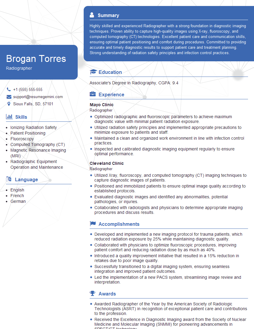
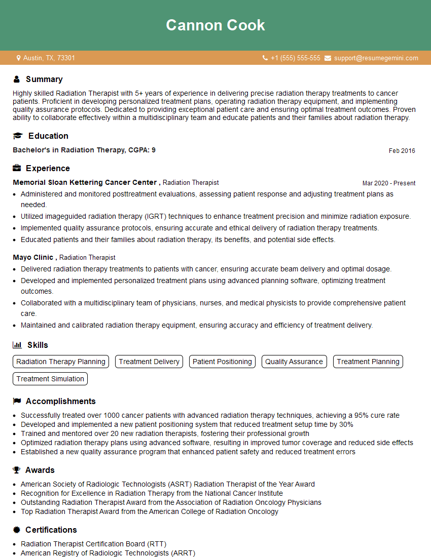
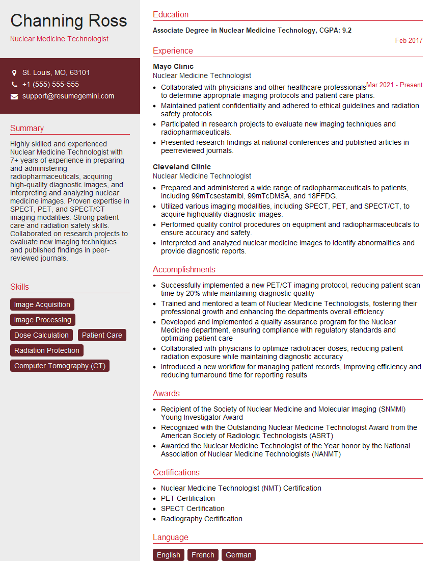
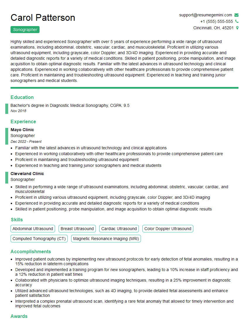
Mastering the skills and knowledge of a Radiology Technician opens doors to a rewarding career with excellent growth potential, offering opportunities for specialization and advancement within the healthcare field. To significantly boost your job prospects, creating a strong, ATS-friendly resume is essential. ResumeGemini is a trusted resource to help you build a professional resume that highlights your skills and experience effectively. Examples of resumes tailored specifically to Radiology Technician positions are available to help guide you. Invest time in crafting a compelling resume – it’s your first impression on potential employers.
Explore more articles
Users Rating of Our Blogs
Share Your Experience
We value your feedback! Please rate our content and share your thoughts (optional).
What Readers Say About Our Blog
Hi, I’m Jay, we have a few potential clients that are interested in your services, thought you might be a good fit. I’d love to talk about the details, when do you have time to talk?
Best,
Jay
Founder | CEO
