Unlock your full potential by mastering the most common Histopathology Technician interview questions. This blog offers a deep dive into the critical topics, ensuring you’re not only prepared to answer but to excel. With these insights, you’ll approach your interview with clarity and confidence.
Questions Asked in Histopathology Technician Interview
Q 1. Describe the process of tissue fixation and its importance in histopathology.
Tissue fixation is the crucial first step in histopathology, where tissue samples are preserved to prevent decomposition and maintain their structural integrity. It’s like taking a snapshot of the tissue’s architecture at a specific moment in time. We achieve this by using fixatives, which are chemical solutions that cross-link proteins and other cellular components, halting autolysis (self-digestion) and putrefaction (bacterial decomposition).
The most common fixative is formalin (10% neutral buffered formaldehyde), which is excellent at preserving morphology. However, other fixatives like Bouin’s solution (picric acid, formaldehyde, and acetic acid) or glutaraldehyde are used for specific applications, such as preserving fine details or certain types of tissue. The choice of fixative depends on the tissue type, the type of staining planned, and the specific information we want to obtain. Inadequate fixation is a major source of artifacts and can compromise the diagnostic accuracy of the specimen. For instance, poorly fixed tissue might show shrinkage, distortion, or loss of cellular detail. Proper fixation ensures that the tissue is ready for subsequent processing steps, maintaining its quality for accurate diagnosis.
Q 2. Explain the different types of tissue embedding methods and their applications.
Tissue embedding is the process of infiltrating the tissue with a supporting medium, usually paraffin wax, to provide structural support for sectioning. Imagine it as creating a mold for the tissue, which will be ‘carved’ later into thin sections for viewing under a microscope. The process typically involves several steps:
- Dehydration: Removing water from the tissue using graded alcohols (e.g., 70%, 95%, 100%).
- Clearing: Replacing alcohol with a solvent miscible with both alcohol and the embedding medium (e.g., xylene or limonene). This process makes the tissue transparent.
- Infiltration: Embedding the tissue in molten paraffin wax. The wax replaces the clearing agent.
- Embedding: Orienting the tissue within a mold filled with paraffin wax and allowing it to solidify. Proper orientation is critical for obtaining sections of the desired area.
While paraffin wax is the most common embedding medium, other methods exist, like freezing (cryoembedding) for rapid processing of tissues needing minimal processing to maintain delicate antigens, or resin embedding for electron microscopy, offering better resolution than paraffin.
The choice of embedding method directly impacts the quality of the final sections and depends heavily on the tissue’s properties and the intended microscopic examination. Cryoembedding, for instance, is quicker but may result in ice crystal formation, whereas resin embedding provides superior resolution but is more time-consuming.
Q 3. What are the common artifacts encountered during tissue processing and how can they be minimized?
Many artifacts can occur during tissue processing, impacting the quality of the final slides and potentially leading to misinterpretations. Common artifacts include:
- Shrinkage and distortion: Caused by inadequate fixation or processing. This can lead to misinterpretation of tissue architecture.
- Precipitation of fixative: Crystals of fixative can obscure tissue details. This can often be due to using overly concentrated fixative or improper washing steps.
- Air bubbles: Trapped air during processing can create spaces in the tissue section.
- Folding and tears: These can occur during embedding or sectioning.
- Over- or under-staining: Improper staining techniques can result in poorly visualized tissue.
Minimizing these artifacts requires meticulous attention to detail at every stage. Proper fixation, careful tissue handling, diligent monitoring of processing times and temperatures, and use of appropriate embedding techniques and quality reagents are all key to preventing artifacts. For instance, gentle handling of tissues during processing, the use of quality reagents, and carefully monitored processing times and temperatures greatly decrease the chances of tears or shrinkage.
Q 4. Describe the principles of microtomy and the different types of microtomes used.
Microtomy is the process of cutting extremely thin sections (slices) of embedded tissue, typically 3-5 micrometers thick, suitable for microscopic examination. It’s a delicate process requiring precision and skill. The principle is simple: a sharp blade is used to cut through the tissue block, producing uniform, thin sections. These sections are then mounted onto glass slides for staining and microscopic analysis. Think of it like slicing a very thin layer off a loaf of bread.
Several types of microtomes exist, each with its own application:
- Rotary microtome: The most common type, using a rotating wheel to advance the tissue block into the blade. It’s versatile and used for paraffin-embedded tissues.
- Sliding microtome: The tissue block remains stationary, while the knife slides across it. Useful for cutting larger or delicate specimens.
- Freezing microtome: Used for cutting frozen tissue, which does not require the dehydration and embedding steps. It’s faster but may produce slightly less crisp sections.
- Ultramicrotome: Used for producing extremely thin sections (nanometers) for electron microscopy.
The choice of microtome depends on the type of tissue, the embedding method, and the desired section thickness.
Q 5. How do you ensure the quality of stained slides?
Ensuring the quality of stained slides is paramount for accurate diagnosis. This involves multiple steps:
- Proper tissue processing: As discussed earlier, minimizing artifacts during processing directly impacts slide quality.
- Careful sectioning: Producing uniform, wrinkle-free sections with the microtome.
- Clean slides: Using clean, grease-free slides to prevent detachment of sections.
- Appropriate staining techniques: Using standardized protocols and quality reagents to ensure consistent and optimal staining.
- Proper mounting: Using a mounting medium that preserves the stained sections and prevents fading.
- Quality control: Regularly checking the staining quality and addressing any inconsistencies. This can involve using control slides with known results.
For example, a common quality control measure is to include a control slide stained with known positive and negative tissue samples in each staining batch to monitor the staining quality and consistency. This ensures that the staining process is working correctly and provides confidence in the results.
Q 6. Explain the process of Hematoxylin and Eosin (H&E) staining.
Hematoxylin and eosin (H&E) staining is the most common staining method in histopathology, providing excellent morphological detail. It’s a two-step process:
- Hematoxylin staining: Hematoxylin, a basic dye, stains acidic components of the cell (like DNA and RNA in the nucleus) a bluish-purple color. Think of it as highlighting the cell’s ‘control center’.
- Eosin staining: Eosin, an acidic dye, stains basic components of the cell (like cytoplasm and extracellular matrix) a pinkish-red color. This provides contrast to the nucleus.
The process involves deparaffinizing (removing paraffin wax), rehydrating (returning water to the tissue), staining with hematoxylin, washing, staining with eosin, dehydrating, clearing, and mounting. Each step must be carefully controlled to achieve optimal staining. The result is a beautifully stained slide where cell nuclei stand out sharply against the background of the cytoplasm. A well-performed H&E stain is the foundation for most histopathological diagnoses.
Q 7. Describe the different types of special stains used in histopathology and their applications.
Special stains are used to highlight specific tissue components that are not well visualized with H&E staining. They are invaluable tools used to confirm diagnoses and provide additional diagnostic information. Examples include:
- Periodic acid-Schiff (PAS): Stains carbohydrates and glycoproteins, often used to detect fungi, glycogen, and basement membranes. It provides a vibrant magenta color.
- Masson’s trichrome: Differentiates collagen from other tissues, useful in assessing fibrosis.
- Silver stains: Used to highlight reticulin fibers, nerve fibers, and microorganisms (e.g., fungi).
- Immunohistochemistry (IHC): Uses antibodies to detect specific proteins or antigens in tissues, allowing for the identification of specific cell types or markers of disease. The technique involves the use of labeled antibodies which binds to specific antigens in the tissue and are then visualized using colorimetric methods.
- Oil Red O: Stains lipids and is used in the detection of fatty changes.
The choice of special stain depends on the suspected diagnosis and the information needed. For instance, a PAS stain might be used to confirm the presence of fungi in a suspected fungal infection, while IHC might be used to detect tumor markers in a suspected cancer sample. Special stains add a layer of detail and accuracy to the histopathological diagnosis, providing the pathologist with critical information that would be otherwise unobtainable with the routine H&E stain.
Q 8. Explain the principles of immunohistochemistry (IHC) and its applications.
Immunohistochemistry (IHC) is a powerful laboratory technique that uses antibodies to identify specific proteins or antigens within tissue samples. Think of it like a highly specific detective searching for a particular criminal (protein) within a crowded city (tissue sample). The principle relies on the highly specific binding between an antibody and its target antigen. We add a labeled antibody (e.g., with an enzyme that produces color or a fluorescent molecule) that binds to the target antigen in the tissue. This allows us to visualize the location and quantity of the antigen within the tissue section under a microscope.
Applications of IHC are vast and include:
- Cancer diagnosis and prognosis: Identifying specific markers associated with different cancer types helps determine the type of cancer, its aggressiveness, and potential response to treatment. For example, ER/PR/HER2 testing in breast cancer guides treatment strategies.
- Infectious disease diagnosis: Detecting specific pathogens within infected tissue allows for precise identification and confirmation of infection.
- Autoimmune disease diagnosis: Identifying autoantibodies or immune cell infiltrates in tissues affected by autoimmune diseases aids in diagnosis and monitoring.
- Neuropathology: IHC helps diagnose neurological conditions by identifying specific proteins associated with neuronal damage or neurodegenerative diseases.
- Research: IHC is invaluable for research into gene expression, protein localization, and cell signaling pathways.
Q 9. What are the safety precautions to be taken while handling hazardous chemicals in the histopathology laboratory?
Safety in a histopathology lab is paramount, especially when handling hazardous chemicals like formalin, xylene, and various stains. Our lab strictly adheres to a comprehensive safety protocol. This includes:
- Personal Protective Equipment (PPE): Mandatory use of lab coats, gloves (nitrile or similar), safety glasses, and sometimes respirators depending on the procedure. We change gloves frequently and dispose of them appropriately.
- Chemical Handling: Proper handling and storage of chemicals in designated cabinets with appropriate labels and safety data sheets (SDS) readily available. We always follow the SDS instructions meticulously.
- Spill Procedures: We have clear protocols for handling chemical spills, including the appropriate neutralizing agents and cleanup methods. Training on spill response is mandatory.
- Fume Hoods: All procedures involving volatile chemicals are performed under fume hoods to prevent inhalation of harmful vapors.
- Waste Disposal: Proper disposal of hazardous waste according to local and national regulations. This involves separating different types of waste (e.g., formalin waste, sharps, etc.) into designated containers.
- Regular Safety Training: Regular safety training and refresher courses ensure that all staff are aware of safety protocols and emergency procedures. We have regular safety meetings and drills.
We treat every chemical with respect and always prioritize safety. A single lapse in procedure can have serious consequences.
Q 10. How do you identify and address potential problems during tissue processing?
Identifying and addressing problems during tissue processing is crucial for producing high-quality slides. Problems can arise at any stage, from tissue fixation to embedding. I use a systematic approach. For instance:
- Inadequate Fixation: Poorly fixed tissues show artifacts such as poor morphology or loss of antigenicity. We address this by checking the fixation time, formalin concentration, and tissue size. If the tissue is inadequately fixed, we may need to refix it.
- Processing Artifacts: These include tissue shrinkage, cracking, or poor tissue infiltration. Checking the processing schedule and reagent quality is crucial. We might need to adjust the processing times or replace reagents if necessary.
- Embedding Issues: Improper orientation or incomplete infiltration of paraffin can lead to difficult sectioning. Careful attention to tissue orientation during embedding is key. Re-embedding may be necessary for optimal sectioning.
- Sectioning Difficulties: Problems such as tearing or compression during sectioning can be caused by dull blades, improper adjustments on the microtome, or hardness of the tissue. We sharpen or replace the blades regularly and adjust the microtome settings as needed. Sometimes using different embedding techniques helps.
Troubleshooting involves careful observation, methodical investigation, and systematic problem-solving. Maintaining meticulous records helps trace the source of problems and ensures that corrective actions are effective.
Q 11. Describe your experience with quality control measures in a histopathology laboratory.
Quality control (QC) is an integral part of our histopathology workflow. We utilize several methods to ensure high quality slides and reliable results:
- Internal Controls: Each batch of slides includes internal positive and negative controls to validate staining techniques and ensure reagents are functioning correctly. This provides immediate feedback on staining quality.
- External Quality Assurance (EQA): Participation in EQA programs allows for comparison of our results with other labs, highlighting areas for improvement and ensuring consistency in our techniques.
- Regular Equipment Maintenance: Routine maintenance of equipment like microtomes, stainers, and embedding stations prevents malfunctions and ensures high quality results. We maintain detailed logs for each piece of equipment.
- Reagent QC: Regular checking of reagent quality and expiration dates prevents the use of degraded reagents that can lead to poor staining or artifacts. I often perform test stainings with new batches of reagents.
- Slide Review: Regular review of slides by senior pathologists and histotechnologists to identify any inconsistencies and address issues in staining quality and tissue morphology.
Our commitment to QC ensures the reliability of our results, which is critical for accurate diagnosis and patient care.
Q 12. Explain your experience with maintaining and troubleshooting histopathology equipment.
I have extensive experience maintaining and troubleshooting various histopathology equipment, including microtomes, tissue processors, embedding centers, stainers, and coverslippers. This involves:
- Preventive Maintenance: Performing routine maintenance according to manufacturer’s instructions, including cleaning, lubrication, and calibration of instruments. We maintain detailed maintenance logs for each equipment.
- Troubleshooting: Identifying and resolving equipment malfunctions using troubleshooting guides and contacting manufacturers for technical support when necessary. Experience has enabled me to solve most issues independently.
- Calibration: Regularly calibrating equipment to ensure accuracy and precision. This is especially important for microtomes to obtain uniform section thickness.
- Software Updates: Keeping the software of automated instruments updated to optimize performance and address any software bugs. I am proficient in the software used by many of our instruments.
- Safety Checks: Conducting safety checks on equipment regularly to identify potential hazards and prevent accidents. This includes checking electrical connections, gas lines (if applicable), and safety features.
My proactive approach to equipment maintenance minimizes downtime and ensures efficient lab operation, leading to a consistent supply of high-quality slides.
Q 13. How do you handle discrepancies in slide preparation and staining?
Discrepancies in slide preparation and staining can stem from various issues. A systematic approach to identify the source of the problem is crucial. My process involves:
- Careful Examination: First, I meticulously examine the affected slides to pinpoint the nature of the discrepancy. Is it a staining issue, a sectioning problem, or a tissue processing artifact?
- Reviewing the Workflow: I trace the slides back through the processing steps, examining the records to identify any deviations from standard protocols or potential errors at each stage.
- Reagent Checks: If the discrepancy points to a staining issue, I check the quality and concentration of the reagents used, as well as their expiration dates. Test stains might be performed.
- Equipment Evaluation: I assess the condition and calibration of the equipment used in the relevant steps (e.g., microtome, stainer). Malfunctioning equipment can be a source of inconsistencies.
- Communication and Consultation: When necessary, I consult with senior histotechnologists and pathologists to discuss the issue and determine the best course of action. This ensures a collaborative and informed approach.
The goal is not just to fix the immediate problem but also to identify and correct the root cause to prevent recurrence. Detailed documentation ensures we learn from each instance.
Q 14. Describe your experience with different types of microscopes and their applications.
My experience encompasses various types of microscopes, each suited to different applications in histopathology:
- Brightfield Microscopes: These are the workhorse of the histopathology lab, used for routine examination of H&E-stained slides. I’m proficient in using these for evaluating tissue morphology, cellular architecture, and identifying various histological features.
- Fluorescence Microscopes: Used for visualizing fluorescently labeled antibodies or other fluorescent probes in IHC or immunofluorescence studies. I’m familiar with different excitation and emission filters needed for different fluorophores.
- Polarizing Microscopes: These are valuable for detecting birefringent structures, like amyloid deposits or crystals, which display unique optical properties under polarized light. I have used this for specific diagnostic purposes.
- Digital Microscopes: These microscopes capture images digitally, which are useful for image analysis, telepathology, and archiving. My experience includes using these systems for case presentations and remote consultations.
Understanding the capabilities and limitations of each microscope type ensures that the most appropriate instrument is selected for each task, improving both the quality and efficiency of diagnostic analysis.
Q 15. How familiar are you with digital pathology and image analysis?
Digital pathology and image analysis are integral parts of modern histopathology. My familiarity extends to utilizing whole-slide imaging (WSI) systems for viewing and analyzing digitized tissue sections. I’m proficient in using various image analysis software to quantify features like cellular density, nuclear size, and staining intensity. This includes experience with both commercially available software packages and open-source platforms. For instance, I’ve used Aperio’s image analysis software for quantifying tumor cell populations in breast cancer specimens and ImageJ for measuring the area of specific tissue structures. This experience allows for more objective and reproducible results compared to traditional microscopy, streamlining the diagnostic process and improving efficiency.
Furthermore, I understand the importance of data management within digital pathology workflows, including image storage, retrieval, and annotation. I’m familiar with different image formats (e.g., TIFF, SVS) and archiving strategies to ensure data integrity and accessibility.
Career Expert Tips:
- Ace those interviews! Prepare effectively by reviewing the Top 50 Most Common Interview Questions on ResumeGemini.
- Navigate your job search with confidence! Explore a wide range of Career Tips on ResumeGemini. Learn about common challenges and recommendations to overcome them.
- Craft the perfect resume! Master the Art of Resume Writing with ResumeGemini’s guide. Showcase your unique qualifications and achievements effectively.
- Don’t miss out on holiday savings! Build your dream resume with ResumeGemini’s ATS optimized templates.
Q 16. Explain your experience with maintaining laboratory records and documentation.
Maintaining accurate and complete laboratory records is paramount in histopathology. My experience encompasses meticulously documenting every step of the process, from specimen accessioning and grossing to tissue processing, embedding, sectioning, staining, and slide archiving. This includes detailed record-keeping in laboratory information systems (LIS) and maintaining physical logs. For example, I consistently document specimen identification numbers, fixative used, processing times, staining protocols, and any unusual findings during tissue handling. I am also familiar with quality control procedures and documentation, ensuring compliance with all relevant regulations and standards. Maintaining these records ensures traceability, accuracy, and efficient retrieval of information when necessary, which is crucial for both diagnostic purposes and quality assurance.
Q 17. How do you prioritize tasks and manage your workload in a busy histopathology lab?
Prioritization and workload management are critical skills in a busy histopathology lab. I utilize a combination of techniques to ensure efficient workflow. I employ a system of prioritizing tasks based on urgency and clinical need. Stat cases, for example, always take precedence, followed by urgent surgical pathology cases. I utilize a task management system – often a combination of to-do lists and the laboratory information system (LIS) – to keep track of assignments and deadlines. I also proactively anticipate potential bottlenecks and proactively address them. For instance, if I notice a large influx of biopsies scheduled for the same day, I will adjust my workflow to ensure timely completion without compromising quality. Effective communication with colleagues is also key; collaborating on tasks and sharing workload helps to maintain a smooth and efficient workflow.
Q 18. Describe your experience with working independently and as part of a team.
I’m comfortable working both independently and as part of a team. Working independently requires strong organizational skills, attention to detail, and the ability to manage one’s time effectively. I thrive in a collaborative environment as well. In a team setting, I value open communication, mutual respect, and the ability to share expertise and knowledge with my colleagues. I’ve regularly assisted less experienced technicians, providing training and guidance on various procedures. In one instance, I collaborated with a pathologist and fellow technicians to troubleshoot a staining issue, ultimately identifying and resolving the root cause and implementing a new quality control measure to prevent future recurrences. Successfully handling both independent and collaborative work scenarios has made me a well-rounded and valuable member of the histopathology team.
Q 19. How do you handle stressful situations in the laboratory?
Stressful situations can arise in a histopathology lab, such as urgent requests, equipment malfunctions, or unexpected sample volume. My approach is to remain calm, prioritize tasks based on urgency, and focus on efficient problem-solving. For instance, if a critical piece of equipment malfunctions, I would immediately notify the appropriate personnel, document the issue, and implement contingency plans to minimize disruption to the workflow. Deep breaths and focusing on the next immediate step are helpful techniques for managing the stress. I also value teamwork in these instances, seeking help and support from my colleagues to resolve the issue quickly and effectively.
Q 20. What are your strengths and weaknesses as a histopathology technician?
My strengths include meticulous attention to detail, a strong understanding of histotechnological principles, and proficiency in various staining techniques. I am highly organized, efficient, and capable of managing multiple tasks simultaneously. One area I am working on is improving my proficiency with advanced image analysis software; while I possess a solid foundation, continuous learning in this rapidly evolving field is essential. I actively seek out opportunities to expand my skills in this area through online courses and workshops.
Q 21. Describe your experience with different types of tissue samples.
My experience encompasses a wide range of tissue types, including but not limited to biopsies from various organs (liver, kidney, lung, etc.), surgical resection specimens, bone marrow aspirates, and cytology samples. I’m familiar with the specific processing requirements for different tissues, taking into account factors such as tissue hardness, fragility, and fixation protocols to optimize tissue preservation and stain quality. For example, I understand the need for decalcification procedures for bone samples and the specific handling techniques required for delicate tissues like brain biopsies. This breadth of experience ensures I can effectively process and prepare a wide variety of samples for accurate microscopic examination.
Q 22. How do you maintain a clean and organized work environment?
Maintaining a clean and organized histopathology lab is paramount for accurate results, efficient workflow, and safety. It’s not just about tidiness; it’s about preventing contamination and ensuring the integrity of samples. My approach is multifaceted:
- Daily Cleanup: I meticulously clean my work station after each procedure, wiping down surfaces with appropriate disinfectants (e.g., 10% bleach solution for grossing areas, 70% ethanol for embedding stations), disposing of sharps and biohazardous waste according to protocol. This prevents cross-contamination between samples.
- Regular Deep Cleaning: Participating in scheduled deep cleans of the entire lab, including equipment like microtomes and embedding centers. This involves thorough cleaning and disinfection of all surfaces and equipment, including attention to hard-to-reach areas.
- Organized Storage: Maintaining a systematic organization of reagents, stains, and other supplies using a FIFO (First-In, First-Out) system. This ensures that older materials are used first to prevent expiry issues and reduces the risk of misidentification.
- Preventative Maintenance: Reporting any equipment malfunctions or maintenance needs immediately. This prevents small problems from becoming larger ones, ensuring the continued proper functioning of essential equipment.
- Proper Waste Disposal: Strict adherence to all biohazard and chemical waste disposal protocols, meticulously labeling and separating different waste types. This is crucial for environmental safety and compliance.
For example, I once noticed a small leak in a reagent bottle during routine cleaning. Immediate reporting prevented a larger spill and potential contamination of other reagents and samples. Proactive maintenance is as important as regular cleaning.
Q 23. What are the ethical considerations related to histopathology practice?
Ethical considerations in histopathology are fundamental to maintaining patient trust and ensuring accurate diagnoses. These include:
- Accuracy and Integrity: Maintaining the highest standards of accuracy in all aspects of tissue processing, sectioning, staining, and reporting. Any deviation from established protocols must be clearly documented and justified.
- Confidentiality: Protecting patient information from unauthorized access or disclosure. This includes adhering to HIPAA regulations and maintaining strict control over patient data, both digital and physical.
- Professionalism: Maintaining professional conduct at all times, avoiding conflicts of interest and adhering to the ethical guidelines set forth by professional organizations like the American Society for Clinical Pathology (ASCP).
- Transparency: Openly communicating any potential errors or limitations in the diagnostic process to the pathologist. This ensures that the pathologist has all the information needed to make an accurate diagnosis.
- Continuing Education: Staying current with advances in histopathology techniques and diagnostic standards. This is essential for providing high-quality services and making informed decisions.
For instance, if I observe a potential error in my own work, I would immediately report it to my supervisor and work collaboratively to find a solution. Ethical conduct means taking responsibility and ensuring patient safety above all else.
Q 24. How do you ensure patient confidentiality in a histopathology lab?
Patient confidentiality is a cornerstone of ethical histopathology practice. In our lab, we maintain confidentiality through several key strategies:
- Strict Accessioning Procedures: Using unique identifiers for each sample, avoiding the use of patient names or other identifying information in the laboratory workflow. Samples are tracked using accession numbers only.
- Secure Data Management: Storing all patient data (both physical and electronic) in secure, password-protected systems. Access is restricted to authorized personnel only.
- HIPAA Compliance: Strict adherence to HIPAA regulations concerning the storage, access, and disposal of Protected Health Information (PHI). This includes training on HIPAA guidelines and regular audits to ensure compliance.
- Secure Disposal of Materials: Proper disposal of all patient samples and associated documents following established protocols. This involves shredding or incineration of documents containing PHI and proper autoclaving or other decontamination methods for tissue samples before disposal.
- Data Encryption: Utilizing encrypted data storage and transmission to prevent unauthorized access even if data is intercepted.
We treat every piece of patient information as highly sensitive, understanding that a breach could have serious consequences. For instance, any requests for patient information are processed through official channels only, with appropriate authorization and documentation.
Q 25. Explain your understanding of relevant health and safety regulations.
Understanding and adhering to health and safety regulations is critical in histopathology. This involves a thorough knowledge of:
- OSHA Regulations: Compliance with Occupational Safety and Health Administration (OSHA) standards concerning the handling of hazardous materials (formaldehyde, xylene, etc.), proper use of personal protective equipment (PPE), and waste disposal.
- CLIA Regulations: Adherence to Clinical Laboratory Improvement Amendments (CLIA) regulations which ensure the quality and accuracy of laboratory testing.
- CAP Accreditation: Understanding and meeting the standards of the College of American Pathologists (CAP) accreditation, which sets high standards for laboratory practices and quality control.
- Biosafety Levels: Knowing and following the appropriate biosafety level (BSL) precautions when handling potentially infectious materials.
- Chemical Safety: Proper handling, storage, and disposal of chemicals following Safety Data Sheets (SDS) guidelines. This includes understanding the hazards associated with each chemical and taking appropriate precautions.
For instance, before handling formaldehyde, I always wear appropriate PPE, including gloves, lab coat, and eye protection, and ensure the work area is well-ventilated. Failure to comply with these regulations can lead to serious health risks and legal consequences.
Q 26. Describe your experience with continuing professional development in histopathology.
Continuing professional development is crucial for remaining at the forefront of histopathology advancements. My commitment to CPD includes:
- ASCP Membership and Resources: Active membership in the American Society for Clinical Pathology (ASCP) provides access to webinars, continuing education courses, and journals which keep me abreast of the latest techniques, technologies and best practices.
- Workshops and Conferences: Attending workshops and conferences on advanced staining techniques, digital pathology, and new diagnostic methods. This provides hands-on experience and networking opportunities.
- Online Courses and Webinars: Taking advantage of online continuing education courses to learn about emerging technologies and best practices in quality control and laboratory management.
- Internal Training: Participating in internal training programs offered by my previous employers to enhance my skills and knowledge in specific areas like immunohistochemistry or molecular diagnostics.
- Mentorship: Actively seeking mentorship from experienced pathologists and histotechnologists to learn from their expertise and gain valuable insights.
Recently, I completed a workshop on advanced immunohistochemistry techniques, which improved my ability to perform complex staining procedures for research and diagnostic purposes. This continuous learning keeps my skills sharp and ensures I can consistently deliver high-quality work.
Q 27. What are your salary expectations for this position?
My salary expectations are commensurate with my experience and skills, and in line with the industry standard for a Histopathology Technician with my qualifications in this geographic area. I am open to discussing a competitive salary range based on the specifics of the position and benefits package.
Q 28. Do you have any questions for me?
Yes, I do have a few questions. First, could you describe the team dynamics and mentorship opportunities within the laboratory? Secondly, what are the opportunities for professional growth and advancement within the organization? Finally, can you elaborate on the specific technologies and equipment used in your histopathology laboratory?
Key Topics to Learn for Histopathology Technician Interview
- Tissue Processing: Understand the entire process from tissue fixation and embedding to microtomy and slide preparation. Consider the impact of different processing techniques on tissue morphology and diagnostic accuracy.
- Microscopy and Staining Techniques: Master the principles of light microscopy, including identifying artifacts and troubleshooting common issues. Gain a strong understanding of various staining methods (H&E, special stains) and their applications in diagnosing different diseases.
- Quality Control and Assurance: Familiarize yourself with quality control measures within the histopathology lab, including proficiency testing, instrument maintenance, and adherence to safety protocols. Discuss practical examples of how you’ve ensured quality in your previous experience.
- Specimen Handling and Accessioning: Learn the importance of accurate specimen identification, tracking, and proper handling to maintain patient data integrity and prevent errors. Practice explaining how you’d manage a high-volume workload efficiently.
- Laboratory Safety and Regulations: Demonstrate a strong understanding of relevant safety regulations (OSHA, etc.) and proper handling of hazardous materials common in a histopathology lab. Discuss practical scenarios where safety protocols are paramount.
- Basic Histopathology: Develop a fundamental understanding of common tissue types and their microscopic appearances. Be ready to discuss how microscopic findings relate to clinical diagnoses. Focus on common pathologies related to your area of interest.
- Troubleshooting and Problem-Solving: Be prepared to discuss instances where you’ve identified and resolved technical issues related to tissue processing, staining, or microscopy. Showcase your analytical and problem-solving skills.
Next Steps
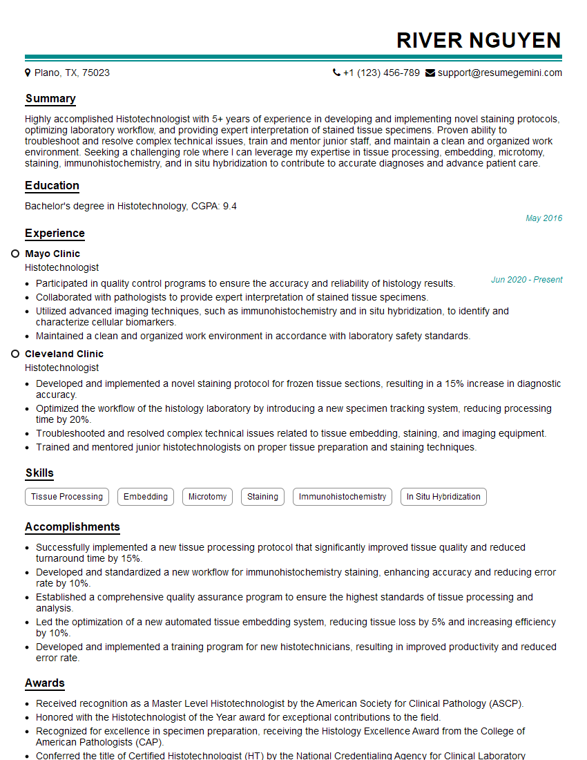
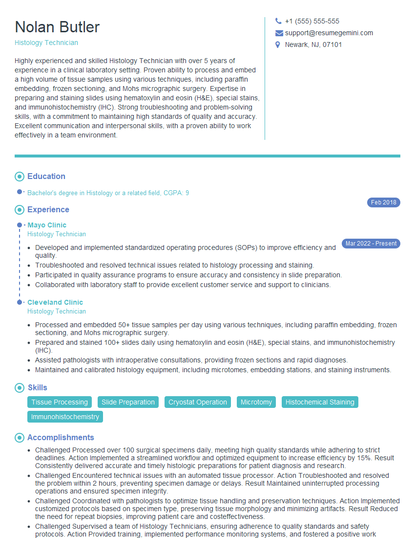
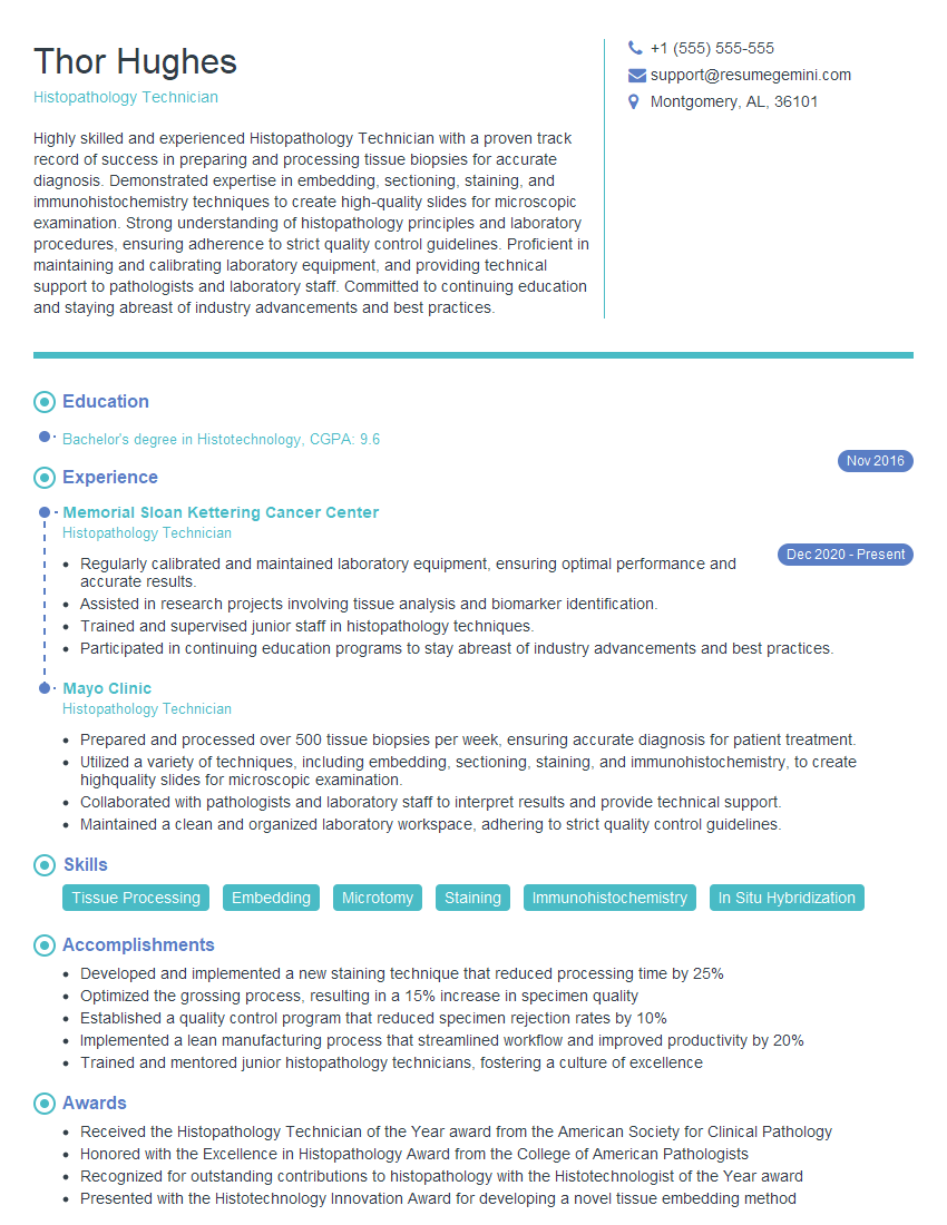
Mastering the skills of a Histopathology Technician opens doors to a rewarding career with excellent growth potential, offering opportunities for specialization and advancement within the medical field. To maximize your job prospects, create a strong, ATS-friendly resume that highlights your skills and experience effectively. ResumeGemini is a trusted resource that can help you build a professional and impactful resume, ensuring your application stands out. Examples of resumes tailored to Histopathology Technician are available to guide you through the process.
Explore more articles
Users Rating of Our Blogs
Share Your Experience
We value your feedback! Please rate our content and share your thoughts (optional).
What Readers Say About Our Blog
Hi, I’m Jay, we have a few potential clients that are interested in your services, thought you might be a good fit. I’d love to talk about the details, when do you have time to talk?
Best,
Jay
Founder | CEO


